
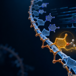


RNA 2′-O-methylation, often abbreviated as Nm, is formed when a methyl group is added to the 2′-hydroxyl group of the ribose sugar. This modification is common in ribosomal RNA, transfer RNA, and small nuclear RNA, and is also found in the 5′ cap structures of many messenger RNAs. Internal Nm sites have additionally been reported in mRNA and other transcript classes, although their distribution and biological significance remain active areas of research.
For researchers, the central challenge is not simply identifying whether Nm exists. The more important question is how to convert a chemical signal into a reliable modification map while separating reproducible evidence from method-specific bias.
How Chemical Treatment Creates a Sequencing Signal
Many Nm sequencing strategies use the different chemical reactivities of methylated and unmethylated ribose groups. In periodate-based workflows, RNA molecules containing an unmodified 2′-hydroxyl group can react differently from ribose residues protected by 2′-O-methylation. After fragmentation, purification, library preparation, and sequencing, these differences can be translated into positional signals.
The resulting reads do not represent a direct photograph of the methyl group. They reflect the combined effects of chemical treatment, RNA structure, fragmentation, ligation, reverse transcription, amplification, sequencing coverage, and computational site calling. This distinction is important because a strong sequencing pattern may indicate a candidate modification site, but it does not automatically establish the modification’s occupancy, biochemical function, or effect on a transcript.
Methods such as Nm-seq and RibOxi-seq were developed to map Nm sites at nucleotide-level resolution. Their chemical principles and enrichment steps are not identical, so results generated by different workflows should be interpreted in the context of the specific assay design. A recent review also highlights the continuing challenges surrounding low-abundance transcripts and internal mRNA Nm sites. Read the review on current challenges in RNA 2′-O-methylation mapping
Why Single-Base Resolution Is Not the Same as Absolute Quantification
Single-base resolution describes how precisely a signal can be assigned to a nucleotide position. It does not necessarily indicate what fraction of molecules in a sample carries the modification at that position.
This difference becomes especially important when comparing biological conditions. A change in signal may result from altered modification occupancy, differences in transcript abundance, unequal representation of RNA classes, or technical variation introduced during sample processing. For this reason, comparisons should consider biological replicates, sequencing coverage, library complexity, and the reproducibility of site calls.
Researchers should also distinguish between a modification map and a functional conclusion. A candidate Nm site may be located within a conserved region, a structured RNA segment, or a transcript associated with a particular pathway. These observations can support hypotheses, but they do not by themselves demonstrate that the modification controls translation, RNA stability, splicing, or another biological process.
Designing the Experiment Around RNA Composition
RNA composition is a major factor in experimental planning. rRNA and tRNA can contribute a substantial fraction of total RNA, while mRNA, lncRNA, and pri-miRNA may be less abundant. The selected RNA input and library strategy should therefore match the biological question.
If the study focuses on abundant structural RNAs, broad total-RNA profiling may provide useful coverage. If the goal is to examine less abundant transcript classes, researchers may need to pay closer attention to RNA enrichment, sequencing depth, library complexity, and the expected signal-to-background relationship.
Sample integrity is equally important. RNA degradation can alter fragment representation and reduce the reliability of downstream mapping. RNA should be carefully quantified, stored under appropriate conditions, and protected from repeated freeze-thaw cycles. These considerations are particularly relevant when the analysis depends on positional enrichment rather than ordinary transcript abundance.
Reading the Data Beyond a List of Sites
A defensible analysis should combine site calling with several layers of quality assessment. Useful checkpoints include:
- read distribution across RNA classes and transcript regions;
- coverage around candidate modification sites;
- replicate consistency;
- library complexity and duplication patterns;
- enrichment or signal distribution after chemical treatment;
- sequence motifs and evolutionary conservation;
- differences between experimental groups.
Functional enrichment analysis can help organize candidate transcripts into biological themes, but it should remain exploratory unless supported by additional evidence. A pathway appearing in an enrichment result does not prove that Nm directly regulates that pathway. Stronger interpretation usually comes from integrating modification data with transcript abundance, RNA structure, translation, or targeted biochemical validation.
When External Analytical Support May Help
A specialized workflow can be useful when a project requires transcriptome-wide Nm mapping across multiple RNA classes, customized bioinformatics analysis, or structured interpretation of site-level results. The 2′-O-Methylation Sequencing service from CD Genomics supports analysis of mRNA, lncRNA, pri-miRNA, tRNA, and rRNA, with workflow stages covering RNA quality assessment, library preparation, sequencing, and downstream data analysis.
Available analysis options may include signal-distribution assessment, methylated transcriptome mapping, site calling, motif analysis, conservation analysis, differential site screening, and functional enrichment. The appropriate analysis depth depends on the RNA composition, experimental design, sample quality, and biological question.
For project discussions, contact Dianna Gellar at contact@cd-genomics.com or +1 631 259 7705. CD Genomics is based in Shirley, NY, USA.
For research use only. Not for use in diagnostic procedures.